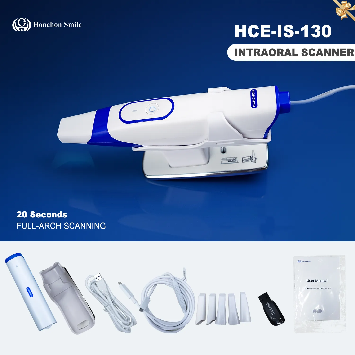
Intraoral scanners (IOS) have become a cornerstone of modern digital dentistry, offering an efficient and patient-friendly alternative to traditional impression techniques. The precision of IOS is directly linked to the clinical success of CAD/CAM restorations, particularly in terms of marginal adaptation and long-term performance. While IOS can achieve accuracy comparable to conventional methods in single-tooth and short-span restorations, challenges remain for long-span and full-arch cases

The accuracy of IOS is generally divided into two components:
Trueness – the degree to which the scanned data represent the actual dentition.
Precision – the consistency and reproducibility of repeated scans under the same conditions.
Both are critical for ensuring reliable digital workflows in restorative and implant dentistry.
Scanner type and design: Performance varies significantly among brands (e.g., TRIOS, iTero, Planmeca) due to different optical and processing technologies
Software version: Continuous updates in acquisition algorithms often enhance data accuracy
Optical system and tip size: Higher resolution and smaller scanner tips improve accessibility in posterior regions
Experience of the operator: Skilled users achieve more consistent outcomes
Scanning path and speed: Recommended manufacturer protocols usually provide the best results, while zig-zag or rotational methods can introduce deviations
Scanning distance: Studies show that scanning at 2.5–5 mm yields higher accuracy than closer or longer distances.
Lighting: Both insufficient and excessive illumination negatively affect scanning. Optimal performance is reported under ambient light between 3,300–10,000 lux .
Environmental stability: Excessive reflections or unstable light conditions may lead to stitching errors .
Tooth position and complexity: Crowding, tilting (>25°), or posterior teeth are associated with reduced accuracy .
Preparation margin location: Subgingival or equigingival margins are harder to capture; gingival retraction significantly improves scan quality .
Implant scan bodies: Their geometry, angulation, and material properties affect accuracy in implant cases .
| Factor | Recommended Strategy |
|---|---|
| Scanner choice | Select reliable brands, ensure latest software updates |
| Scanning protocol | Follow manufacturer’s scanning path, maintain optimal distance |
| Environment | Ensure uniform lighting, avoid direct reflections |
| Preparation design | Favor supragingival margins when possible; use gingival retraction |
| Operator training | Invest in training and standardization of scanning techniques |
The accuracy of intraoral scanners depends on a complex interplay of device-related, operator-dependent, environmental, and patient-specific factors. By recognizing and addressing these variables, clinicians can maximize the reliability of digital impressions and ensure better restorative outcomes.